If you've been scheduled for a scan, you're likely wondering what the technician is actually looking for and how they decide if a nodule is "suspicious." The process is less about a simple "yes or no" regarding cancer and more about calculating a risk score based on the visual characteristics of the nodule. By the end of this guide, you'll understand the markers of risk and the steps taken to ensure an accurate diagnosis.
Why Ultrasound is the First Choice
When a doctor suspects a thyroid issue, they don't start with a CT scan or an MRI. Why? Because Thyroid Ultrasound is significantly more sensitive. While a physical exam might only detect 2% to 21% of nodules, an ultrasound can catch between 19% and 68% of them. It's faster, cheaper, and safer since there's no ionizing radiation involved.
Unlike other imaging, ultrasound allows the radiologist to see the "texture" of the nodule in real-time. They can see if a nodule is filled with fluid (cystic) or if it's a solid mass. They can also use Doppler imaging to check blood flow. If a nodule has significant blood flow in its center, it can sometimes be a red flag for malignancy, whereas blood flow only on the edges is often more typical of benign growths.
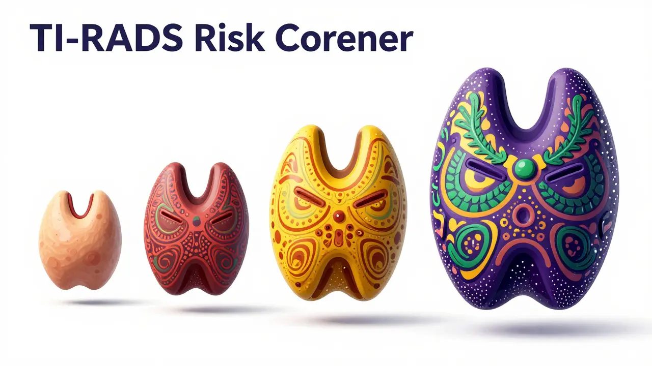
Decoding the TI-RADS Risk Score
Radiologists don't just guess if a nodule looks "weird." They use a standardized system called TI-RADS (Thyroid Imaging Reporting and Data System). This system assigns points based on five specific visual features. The more points a nodule earns, the higher its risk category.
| Category | Points | Estimated Cancer Risk | Typical Action |
|---|---|---|---|
| TR1 (Benign) | 0 | 0.3% | No follow-up needed |
| TR2 (Not Suspicious) | 2 | 1.5% | Usually no follow-up |
| TR3 (Mildly Suspicious) | 3 | 4.8% | Biopsy if > 2.5 cm |
| TR4 (Moderately Suspicious) | 4-6 | 9.1% | Biopsy if > 1.0 cm |
| TR5 (Highly Suspicious) | 7+ | 35% | Biopsy if > 1.0 cm |
So, what exactly are they scoring? They look at composition (is it solid or spongy?), echogenicity (how dark is it compared to the rest of the gland?), shape (is it taller than it is wide?), margins (are the edges smooth or jagged?), and echogenic foci. The last one is a big deal: tiny white dots known as microcalcifications often push a nodule into a higher risk category because they are frequently associated with papillary thyroid cancer.
Ultrasound vs. Other Imaging Methods
You might hear about "nuclear medicine" or "thyroid scans." It's important to distinguish these from ultrasound. A nuclear scan tells us if a nodule is "hot" or "cold." A hot nodule produces thyroid hormone and is almost never cancerous (less than 1% risk). A cold nodule doesn't produce hormone and has about a 15% chance of being malignant. However, a nuclear scan can't tell you what the nodule actually looks like-it only tells you how it functions.
CT scans and MRIs are great for seeing if a tumor has spread to other parts of the neck or if a goiter is growing down behind the breastbone (substernal goiter), but they lack the resolution to see the micro-details needed for initial risk grading. Ultrasound remains the primary tool because it can guide the next critical step: the biopsy.
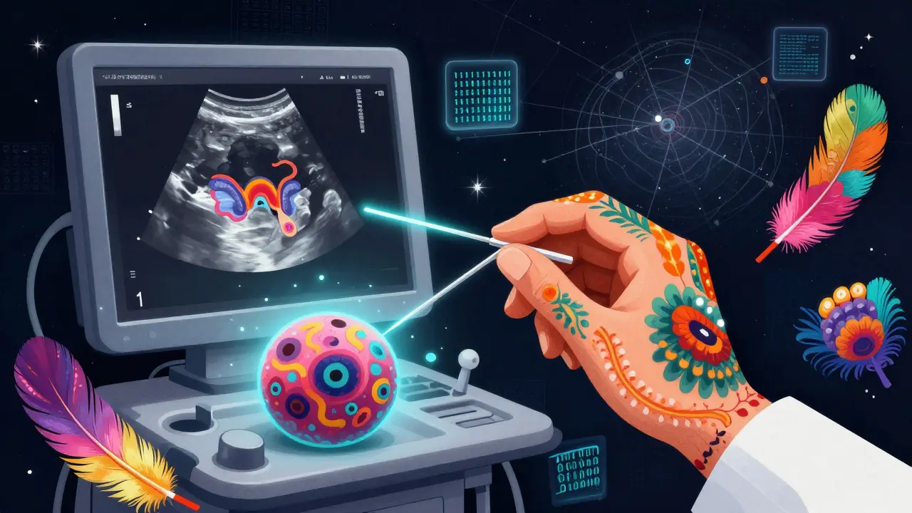
The Path to Diagnosis: From Imaging to Biopsy
If your TI-RADS score is high enough, the next step is usually a Fine-Needle Aspiration (FNA). This is where a very thin needle is used to take a small sample of cells from the nodule. The magic of ultrasound here is that it acts as a GPS. By watching the needle in real-time, the doctor ensures they are hitting the most suspicious part of the nodule. This precision has dropped the rate of "inadequate samples" from 25% down to less than 5%.
But here is the catch: sometimes the biopsy comes back as "indeterminate." This means the pathologist can't tell for sure if the cells are benign or malignant. In these cases, doctors may use molecular testing to look for specific genetic mutations. Even then, ultrasound surveillance is kept in place to monitor any changes in size or appearance over time.
Modern Tech: AI and Beyond
We are entering an era where computers are helping radiologists read these scans. Deep learning models are now being used to analyze nodule aspect ratios and patterns. Some recent studies have shown that AI can boost diagnostic accuracy to over 94%, helping to reduce the a gap between a novice sonographer and an expert radiologist.
There are also newer techniques like elastography, which measures how stiff a nodule is (cancerous tissues are often stiffer than benign ones), and contrast-enhanced ultrasound. While these aren't standard yet, they provide extra layers of evidence when a standard grayscale image leaves the doctor unsure.
What to Expect During and After Your Scan
The procedure itself is straightforward. You'll lie on your back, and a clear gel will be applied to your neck. The sonographer will move a transducer-a handheld probe-across the area. You might feel some pressure, but it's generally painless. The most important thing is to ensure the technician checks the cervical lymph nodes in your neck, as these are critical for staging if cancer is suspected.
Once the scan is over, don't panic if you see "nodules" on your report. As mentioned, they are incredibly common. The key is the TR category. If you are a TR1 or TR2, you can usually breathe easy. If you are TR4 or TR5, it doesn't mean you have cancer; it means the nodule has features that warrant a biopsy to be safe. Many small, highly suspicious nodules (micropapillary cancers smaller than 1 cm) are now managed with "active surveillance"-meaning they are just watched-because the 10-year survival rate for these is over 99%.
Does a thyroid ultrasound definitely find cancer?
No. An ultrasound is a risk assessment tool, not a definitive diagnostic tool. It identifies suspicious features and assigns a risk score (like TI-RADS), but only a biopsy (Fine-Needle Aspiration) or surgical pathology can confirm whether a nodule is actually cancerous.
What is the difference between a "hot" and "cold" nodule?
These terms come from nuclear medicine scans, not ultrasound. A "hot" nodule takes up radioactive iodine and produces hormone; these are rarely malignant. A "cold" nodule does not take up iodine and has a higher risk (around 15%) of being cancer, which is why they are usually followed up with an ultrasound.
Why is "taller-than-wide" shape considered suspicious?
Benign nodules tend to grow along the natural planes of the thyroid tissue, making them wider than they are tall. Malignant tumors often grow aggressively in all directions, including vertically, which creates that "taller-than-wide" appearance on the scan.
How often do I need a follow-up ultrasound?
This depends on your TI-RADS score. Benign nodules may not need any follow-up. Mildly suspicious ones might be checked every 6 to 12 months to see if they grow or change. Your doctor will set a schedule based on the specific features of your nodule.
Is the gel used during an ultrasound safe?
Yes, the ultrasound gel is water-based, hypoallergenic, and safe for all skin types. It's used simply to eliminate air between the probe and your skin, which allows the sound waves to travel into the thyroid gland.
Next Steps and Troubleshooting
If you've just received your results and feel overwhelmed, here is a simple way to handle the next steps:
- For Low Risk (TR1-TR2): Discuss with your doctor if any symptoms (like trouble swallowing) are present. Usually, no further action is needed.
- For Intermediate Risk (TR3): If the nodule is under 2.5 cm, you'll likely just be monitored. If it's larger, ask your doctor if an FNA is appropriate.
- For High Risk (TR4-TR5): If the nodule is over 1 cm, request a referral for a Fine-Needle Aspiration. If you are anxious, ask about the possibility of molecular testing if the biopsy results are indeterminate.
- If you have a "Cold" Nodule: Ensure your ultrasound specifically looks for microcalcifications and the "taller-than-wide" shape, as these are the strongest indicators of risk.